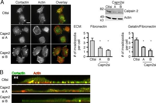
Figure 1.
Calpain is necessary for invadopodia formation. (A) Cell lysates from MTLn3 cells stably expressing control or calpain 2 siRNA were analyzed by Western blotting and probed for calpain 2 and actin as a loading control. MTLn3 cells expressing control or calpain 2si (targets A and B) were cultured on FN-coated glass coverslips and stained with anti-cortactin antibody (green) and rhodamine phalloidin (red). Quantification of cortactin and actin containing invadopodia is expressed as the mean number of invadopodia per cell for FN-coated and gelatin/FN-coated coverslips. Data are mean ± SEM of three independent experiments. *, P < 0.05 compared with control cells. Bar, 10 μm. (B) Confocal images demonstrate actin (red) and cortactin (green) staining at protrusive structures on the ventral cell surface of control or calpain 2 si cells.

