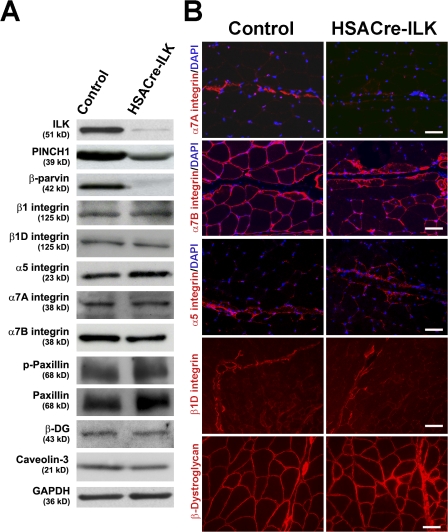
Figure 6.
Expression and distribution of ILK-associated proteins. (A) ILK, Pinch, and parvin are decreased in HSACre-ILK muscle, whereas β1, β1D, α5, α7A, and α7B integrin and phosphopaxillin (p-paxillin), β-dystroglycan (β-DG), and caveolin-3 are not changed. GAPDH was used as the loading control. (B) Immunofluorescence staining of α7A, α7B, α5, and β1D integrins and β-dystroglycan. Note the prominent α7A integrin signals (red) at MTJ of control muscles and the reduction at MTJs of HSACre-ILK muscles. Although α7B integrin (red) is expressed on the sarcolemma of all myofibers in the control, it shows an irregular staining pattern in HSACre-ILK muscle. In contrast to the control muscle, α5 integrin signals are not restricted to the tendon of HSACre-ILK muscles. β1D Integrin and β-dystroglycan signals are comparable between control and HSACre-ILK muscles. Bars: (α7A integrin, α5 integrin, and β-dystroglycan) 40 μm; (α7B integrin) 35 μm; (β1D integrin) 60 μm.

