Figure 7.
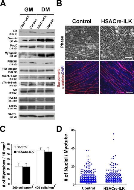
Normal fusion of HSACre-ILK myoblasts. (A) Protein levels of ILK and PINCH1 are dramatically decreased in primary HSACre-ILK myoblasts both in GM and DM. Signals of desmin and myogenin increase and MyoD decreases in response to differentiation in both cell types. The levels of β1D integrin, pSer473-Akt, pThr308-Akt, Akt, pErk1/2, and Erk1/2 are comparable. GAPDH was used as the loading control. (B) Myoblast cells were isolated from postnatal day 2 control and HSACre-ILK hindlimbs, incubated in DM, and evaluated microscopically. Differentiated myoblasts and myotubes were stained with sarcomeric α-actinin antibody (red) and DAPI (blue). (C) Quantification of myotube numbers from control and HSACre-ILK myoblasts plated at different cell densities. Data are expressed as mean ± SD (n = 3; 200 and 400 cells/nm2). (D) Determination of the number of nuclei per fused myotube. No difference between control and HSACre-ILK mice was observed (P = 0.253). Bars: (phase) 50 μm; (sarcomeric α-actinin) 80 μm.

