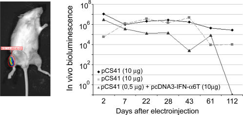
Figure 3. Expression of luciferase, in vivo, after i.m. electroinjection of an expression plasmid.
Left panel: picture of a mouse showing luciferase expression in the tibialis muscle of the right leg. Right panel: follow-up of luciferase expression in vivo (arbitrary units), in two mice electroinjected with 10 µg of plasmid DNA (pCS41) expressing the firefly luciferase gene and in one mouse electroinjected with 0.5 µg of pCS41 plasmid DNA and 10 µg of plasmid DNA expressing IFN-α6T.

