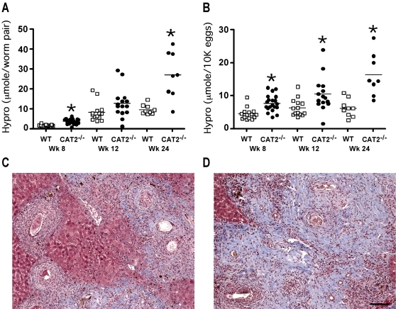
Figure 3. S. mansoni infected CAT2−/− mice develop exacerbated liver fibrosis.
WT C57BL/6 (open squares) and CAT2−/− (filled circles) mice were infected with 30–35 S. mansoni cercariae and sacrificed on wk 8, 12 and 24 post-infection. A. Liver Fibrosis adjusted per worm pair (µmol of hydroxyproline/worm pair). The * symbol denotes significant differences between WT and KO mice at that time point, p<0.05. B. Liver Fibrosis per 10,000 eggs (µmol of hydroxyproline/1×104 eggs). C. Representative liver granulomas stained with Masson's Trichrome (8 wk infected C57BL/6 mouse). D. Representative liver granulomas stained with Masson's Trichrome (8 wk infected CAT2−/− mouse). The bar in panel 3D = 200 microns.

