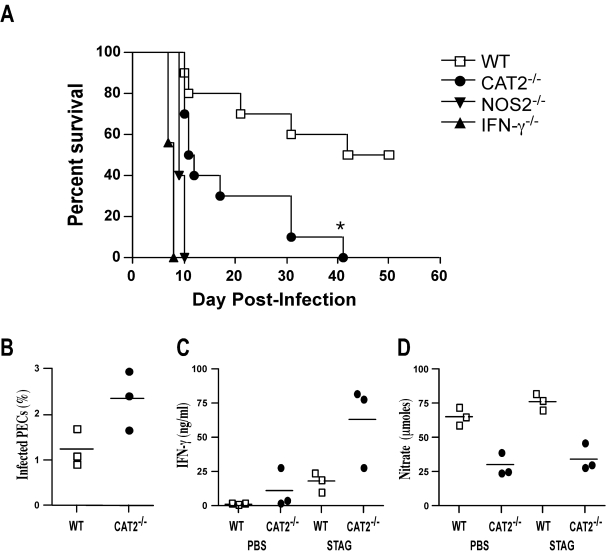
Figure 11. Development of Th1 immunity is compromised in CAT2−/− mice.
A. WT C57BL/6 (open squares), CAT2−/− (filled circles), NOS2−/− (filled inverted triangles) and IFN-γ−/− (filled triangles) mice were infected i.p. with 20 T. gondii cysts, and survival was assessed up to 50 days post-infection. (N = 5/group). B. Peritoneal exudate cells (PECs) were prepared from WT and CAT2−/− mice on day 7 post-infection. The percentage of infected cells was determined microscopically by evaluating a minimum of 700 cells per slide (3 mice/group). C. PECs were placed in culture for 24–48 hr and either left untreated or restimulated with STAG. Culture supernatants were collected and IFN-γ levels were determined by ELISA. D. NO activity was evaluated in the same culture supernatants by measuring nitrate production.

