
Figure 1.
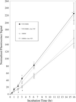
Normalized fluorescence signal of vasoactive intestinal peptide-grafted, quantum dots-encapsulated sterically stabilized mixed phospholipid micelles (VIP-SSMM-QD) and SSMM-QD in human MCF-7 cells in the absence and presence of excess free VIP (30 µM). Each group, n=5; * p<0.05 for VIP-SSMM-QD in comparison to SSMM-QD, and SSMM-QD with excess free VIP; # p<0.05 for VIP-SSMM-QD in comparison to VIP-SSMM with excess free VIP. Total number of cells counted for each time point: 0.5 h, 3477; 1 h, 5310; 2 h, 4405; 3 h, 6419; 6 h, 3970; 16 h, 2266.