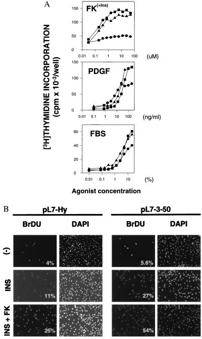
Figure 3.
Rap1b-expressing cells show an increased ability to traverse S phase. (A) [3H]Thymidine incorporation assays indicate that Rap1b- expressing cells (pL7-3-24, ▪ and pL7-3-50, ▴) show an increase in the maximal response, as compared with control (pL7-Hy, •) cells. Dose-responses were performed for fetal bovine serum (FBS), platelet-derived growth factor (PDGF) and forskolin (FK). Forskolin dose-response was performed in the presence of a constant amount of insulin (Ins, 1 μg/ml) and 3-isobutyl-1-methylxanthine (100 μM). (B) Nuclear incorporation of BrdUrd was assayed by indirect immunofluorescence (32). Cells were left untreated (−), or stimulated with insulin (INS, 1 μg/ml) or insulin plus forskolin (INS+FK, 1 μg/ml and 10 μM, respectively) in the presence of 100 μM 3-isobutyl-1-methylxanthine. Total nuclei was visualized by 4′,6-diamidino-2-phenylindole staining, and the results expressed as % BrdUrd/4′,6-diamidino-2-phenylindole. Experiments were done in duplicates and three or four independent fields per sample analyzed and expressed as averages (variation <10%).

