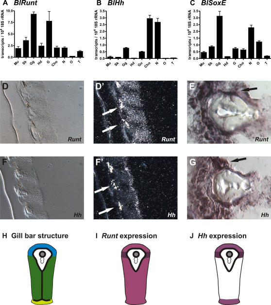
Figure 8. Analysis of Runt, SoxE and Hh gene expression in adult lancelet (B. lanceolatum).
(A–C) Quantification of Runt, SoxE and Hh expression in different tissues. (A) The strongest Runt expression is seen in the gill gut region followed by the gut and skin. (B) Hh is most strongly expressed in the chorda and neural tube followed by the gill gut and gut. (C) SoxE has its strongest expression in the gill gut and neural tube. Mu: Muscle, Sk: Skin, Gg: Gill gut, Hd: Hepatic diverticulum, G: Gut, Cho: Chorda, N: Neural tube, O: Ovaries, T: Testis. (D–G) in situ hybridization for BlRunt and BlHh show high expression in the endoderm and ecotoderm of the gill bars. (D–E) Runt expression. (F–G) Hh expression. (D, F) Bright field images. (D′, F′) Dark field images of radioactive in situ hybridizations. (E, G) Non-radioactive in situ hybridizations. High expression of BlRunt and BlHh was found in a cell population between the endodermal epithelium with cilia and the ectodermal gland epithelium directly adjacent to both sites of the acellular matrix (arrows). (H–J) Schematic drawing of Runt and Hh expression sites in secondary gill bars as shown in (D–G). (H) The gill bar tissue consists of three different single layered epithelia attached to a basal membrane - atrial epithelium (blue), lateral epithelium (dark green) and pharyngeal epithelium (light green). The basal membrane is indicated by the bold black line. The skeletal rod of secondary gill bars contains a skeletal vessel (grey filled circle) that is formed by basal membranes, and does not contain endothelial cells. (I) Runt expression is found throughout the gill bar epithelia (light purple) with strongest expression adjacent to the skeletal rods (dark purple). (J) Hh is expressed at weaker levels in the atrial and pharyngeal epithelium (light purple) and at high levels in the cell population adjacent to the skeletal rods (dark purple).

