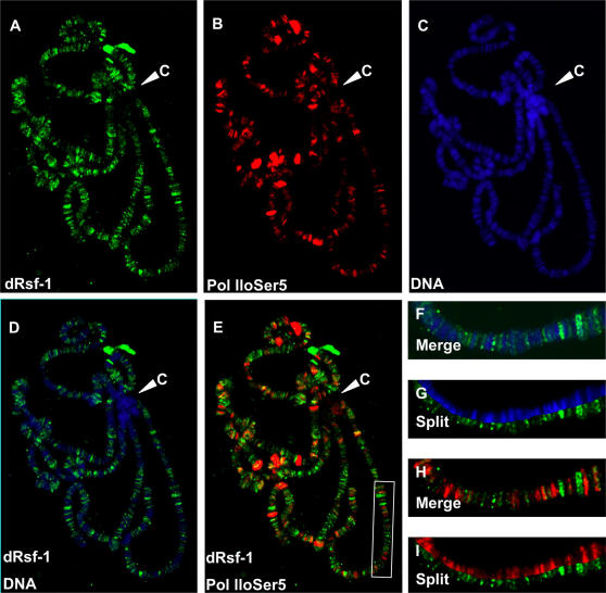
Figure 2. Localization of dRsf-1 protein on polytene chromosomes.
(A–C) Distribution of dRsf-1 protein (green), Ser5-pohosohorylated Pol ll (red) and DNA stained with DAPI (blue) on wild-type polytene chromosomes. Merged and split images are shown in (D,F,G): dRsf-1 and DNA, and in (E,H,I): dRsf-1 and Pol lloser5. dRsf-1 was primarily associated with interband regions, but not transcribed regions. No or very few dRsf-1 were associated with chromocenter (C). (F–I) Higher magnifications of the chromosome (white rectangle in (E)) are shown.

