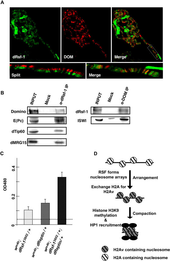
Figure 8. RSF interacts with Tip60 complex.
(A) Distribution of dRsf-1 protein (green) and Domino (red) on wild-type polytene chromosomes. The merged image is shown in the right panel (Merge). Extensive overlap between dRsf-1 and Domino on chromosome 3R is evident in the enlarged merged and split images of the region encompassed by a white rectangle (lower panel). (B) Co-immunoprecipitation of RSF and Domino. Drosophila embryonic nuclear extracts were used for immunoprecipitation using anti-dRsf-1 antibodies (left panel), anti-Domino antibodies (right panel) and control IgG (Mock). The proteins were separated by SDS-PAGE and analyzed by western blotting. Several subunits of the Tip60 complex were co-immunoprecipitated with anti-dRsf-1 antibodies. RSF was co-immunoprecipitated with anti-Domino antibodies, but not with control IgG. Input represents 10% of the starting extract used for the immunoprecipitation. (C) Genetic interaction between dRsf-1 and dReptin. The pigments extracted from male heads of each fly lines were measured at 485 nm. Error bars indicate standard deviations from independent four or three measurements (shown in the graph). Dotted line shows the value for the control fly (wm4h; +/+). (D) Proposed pathway for silent chromatin formation. RSF forms nucleosome arrays and contributes to the exchange of H2A with H2Av in an early step of the silent chromatin formation. These nucleosome arrays are stabilized by the internucleosome interactions through H2Av proteins. This triggers the following steps of the silent chromatin formation such as the recruitment of HP1 and the methylation of H3K9.

