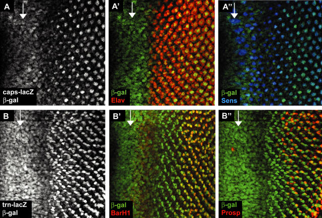
Figure 1. caps and trn expression in the eye.
The arrow in each panel marks the morphogenetic furrow (MF) and anterior is to the left in all images, unless otherwise stated. (A) caps-lacZ expression in 3rd instar eye disc. Staining with anti-β−gal revealed caps-lacZ expression in the furrow and in subsets of cells after the furrow. (A'–A”) Co-staining with anti-Elav (a photoreceptor marker) and anti-Senseless (R8 specific marker) identified the cells eventually expressing high levels of caps-lacZ as photoreceptor R8. (B) trn-lacZ expression in 3rd instar eye disc. Staining with anti-β−gal revealed trn-lacZ expression in the furrow and in a different subset of cells from caps-lacZ after the furrow. (B'–B”) Co-staining with anti-BarH1 (R1 and R6 specific marker) and anti-Prospero (R7 and cone cell marker) identified R1, 6 and 7 as the photoreceptors expressing high levels of trn-lacZ.

