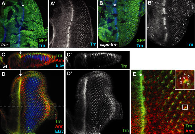
Figure 2. Localisation of Tartan protein in the eye.
(A–A') The Trn antibody recognised endogenous levels of Trn in the 3rd instar eye disc. The signal is absent in clones of trn28.4 null cells (marked by loss of GFP, green), confirming antibody specificity. (B–B') The Trn antibody signal was also lost in capsDel1trn28.4 double null clones, marked by loss of GFP (green). (C–C') Z-section along the A-P axis of a wild type disc stained with anti-Trn. Trn is expressed only in the anterior half of the furrow and mostly in the apical surface of photoreceptors, as well as in some basolateral membranes. (D–D') Apical planar views of the corresponding discs in (C–C'). Trn is expressed in the furrow and in subset of photoreceptors after the furrow. The dashed line indicates the position of the sagittal section in C and C'. (E) Enlarged view of the disc in D. The inset shows an enlarged view of the marked ommatidium with the positions of each photoreceptor labelled. Trn expression is located in the expected positions of R1, 6, 7.

