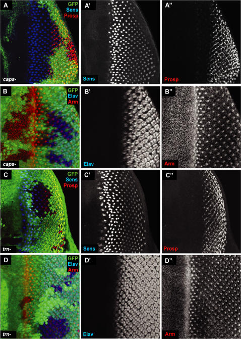
Figure 4. caps and trn single null clones.
(A–B) capspB1 clones in the 3rd instar eye disc. Mutant tissue is marked by lack of GFP (green). Anti-Senseless (Sens), is used to identify R8 cells (A') and anti-Prospero (Prosp) is used to identify R7 and cone cells (A”). Anti-Elav marks all photoreceptor cells (B') and anti-Armadillo (Arm) marks the adherens junctions of cells, thereby outlining all cells (B”). No defects could be detected in capspB1 mutant tissue. (C–D) trn28.4 clones in the 3rd instar eye disc. Mutant tissue is marked by lack of GFP (green). As with capspB1 clones, no defects were observed.

