Figure 5.
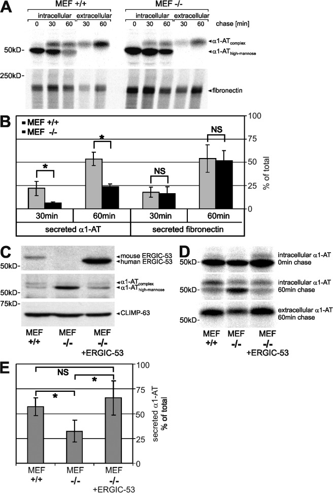
ERGIC-53 is an intracellular transport receptor of α1-AT. α1-AT was transfected into wild-type (+/+) and ERGIC-53 knockout (−/−) MEFs and expressed for 24 h. (A) [35S]methionine pulse chase of α1-AT and endogenous fibronectin. MEFs were labeled with [35S]methionine for 15 min and chased for the indicated times, and α1-AT and fibronectin were recovered by immunoprecipitation from cell lysates (intracellular) and conditioned medium (extracellular) using anti–α1-AT and anti-fibronectin antibodies, respectively. (B) The secreted fraction of α1-AT and fibronectin was quantified as described in Fig. 4 C. (C) α1-AT was coexpressed with human ERGIC-53 in ERGIC-53 knockout MEFs (MEF −/− plus ERGIC-53). Western blotting was performed using polyclonal antibodies against ERGIC-53, α1-AT, and CLIMP-63. (D) [35S]methionine pulse chase of α1-AT. (E) The secreted fraction of α1-AT after a 60-min chase was quantified by densitometric scanning and calculated as in Fig. 4 [extracellular/(intracellular + extracellular)]. Bars represent mean ± SD (n = 3). Results analyzed by paired t test: NS, P > 0.05; *, P < 0.05.

