Figure 5.
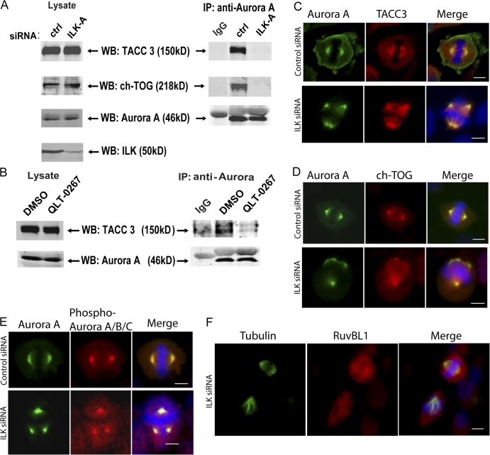
ILK siRNA causes a disruption of Aurora A–TACC3/chTOG interaction. (A) HeLa cells were transfected with control or ILK siRNA and synchronized with nocodazole. Aurora A kinase was immunoprecipitated from cytoskeletal extracts with a monoclonal anti–Aurora A antibody and then Western blotting was performed with polyclonal antibodies against Aurora A, TACC3, and ch-TOG. (B) A similar experiment to A was performed on cells treated with QLT-0267 and a DMSO control. (C–F) Effect of ILK siRNA on localization of ILK-interacting proteins. Hela cells were transfected and stained as indicated. Phospho–Aurora A/B/C staining is presumably phospho–Aurora A rather than Aurora B or C, as it colocalizes with total Aurora A; Aurora B is not found on centrosomes, and Aurora C is only expressed in testis. Bars, ∼10 μm.

