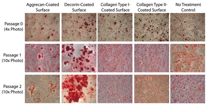
Figure 1. Safranin O staining of TMJ disc cells plated on protein-coated surfaces.
Safranin O/Fast Green staining was used to detect nodule formation on the protein-coated surfaces. At passage 0, all surfaces had both nodules and spindle-shaped cells, and cell shape and GAG production appeared similar on all surfaces. At passage 1, aggrecan-coated and decorin-coated surfaces caused large nodule formations, while collagen type I-coated, collagen type II-coated, and no treatment controls had relatively more spindle-shaped cells. At passage 2, aggrecan-coated and decorin-coated surfaces again caused large nodule formations. Nodules on the aggrecan-coated surface were spread across the well, while nodules on the decorin-coated surface were large and concentrated near the center of the well. Nodule formation was observed on each surface at each passage; however, the size and prevalence of nodules was lower on collagen-coated surfaces and no treatment controls.

