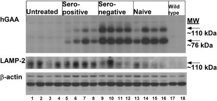
Figure 3. .
Western blot detection of hGAA in cardiac muscle after administration of rhGAA (100 mg/kg). Cardiac muscle was analyzed 3 wk after administration of rhGAA. Three different proteins were detected, hGAA, LAMP-2, and β-actin. β-actin served as a control to indicate equal loading of each lane. Samples were from GAA-KO mice; either untreated, or rhGAA-treated seropositive, seronegative, and naive mice, n=4 for each group; and from C57BL/6 wild-type controls (n=2). Each lane represents an individual mouse.

