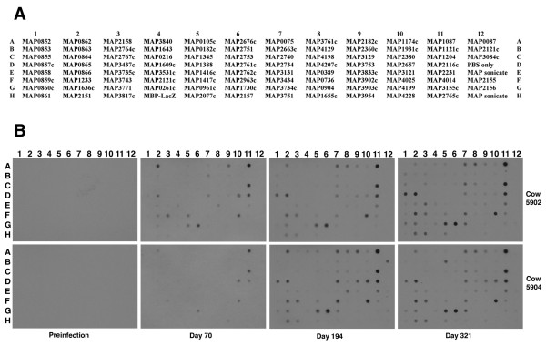
Figure 2.
Use of the dot blot protein array to obtain antibody reactivity profiles of experimentally infected calves. Shown are the spot assignments for the array (A) and dot blot arrays exposed to sera from experimentally infected calves (B). The time point for when each serum sample was collected is indicated in the margin beneath the images. The animal number is listed in the right margin. A whole cell lysate representing a majority of the proteins produced by M. paratuberculosis is spotted in E12 and H12 for all dot blots. Three proteins present on the upper right corner of the array (in column 12) are polyhistidine tagged proteins (MAP0087, MAP2121c and MAP3084c). The remaining 89 spots contain MBP fusion proteins of M. paratuberculosis coding sequences. Note that the MAP2121c coding sequence is represented twice on the array; once as an MBP fusion (spot F4) and also a polyhistidine tagged protein (12B).

