
Fig. 2. δ-Catenin undergoes Akt1-mediated phosphorylation at Thr-454 residue.
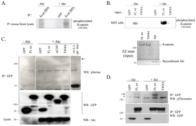
An Akt1 kinase assay was performed as described in Materials and Methods. GFP, mock transfected proteins were used as a negative control in the assay, and GFP-immunocomplex was obtained by immunoprecipitating with GFP antibody. (A) Purified immunocomplex of 1 day post-natal mouse brain lysates with δ-catenin (BD) antibody was used as a substrate for an Akt kinase assay. [γ-32P]-ATP was supplemented with a reaction buffer, and phosphorylation status was detected by autoradiograph. (B) GFP-tagged δ-catenin wt (FL wt) and T454A mutant were transfected in MEF cells, and purified immunocomplex with δ-catenin (BD) antibody was used as a substrate for an Akt kinase assay. [γ-32P]-ATP was supplemented with a reaction buffer, and phosphorylation status was detected by autoradiograph (Upper panel). The level of δ-catenin FL wt and T454A mutant existing in immunocomplex and of recombinant Akt1 were confirmed by EZ staining kit (Bottom panel). (C-D) GFP-tagged full length (FL wt) and mutant δ-catenin (ΔC207 and T454A) were overexpressed in Bosc23 cells, and purified immunocomplex with GFP antibody was used as a substrate for Akt kinase assay. Radio-inactive cold ATP was supplemented with a reaction buffer, and phosphorylation status on serine and threonine residue in the wild type or mutant type δ-catenin was detected using anti-phosphoserine antibody (C) or with anti-phosphothreonine antibody (D) (upper panel). HA-14-3-3 was immunoprecipitated with HA antibody and was used as a positive control (C). An arrow indicates the predicted site (C) or an actual band (D) of phosphorylated δ-catenin, and an asterisk indicates a nonspecific band. The immunocomplexes were sub sequentially reprobed with anti-GFP antibody (middle panels), and the added recombinant Akt1 was also confirmed using anti-Akt antibody (C, bottom panel).