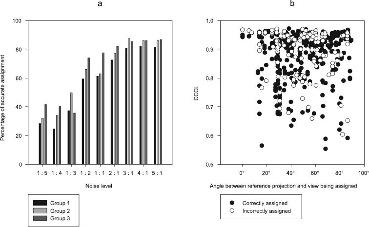
Figure 6.
3D conformational sorting of eIF3-IRES
(a) Conformational variability in 2D averages of eIF3-IRES described by three distinct conformations of IRES bound to eIF3 in a single projection view.
(b) Corresponding 3D reconstructions resulting from the application of the CCCL approach. The blue conformer corresponds closely to that of IRES bound to the 40S ribosome (Spahn et al. 2001).

