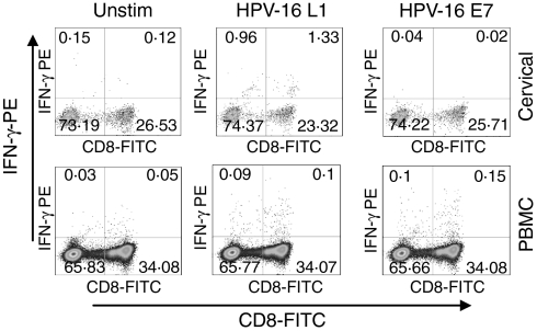
Figure 1.
Representative plots of cervical and blood mononuclear cells from a patient with CIN 2/3 stimulated directly ex vivo with HPV-16 L1 and E7 and stained for intracellular IFN-γ production. IFN-γ production by cervical (upper panel) and blood CD3+ cells (lower panel) either unstimulated or following stimulation with HPV-16 antigens L1 (VLPs) and E7. Each plot was gated on CD3+ cells and then analysed for CD8+ and IFN-γ production.

