Figure 3.
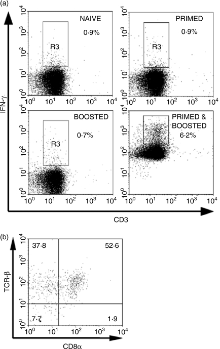
Frequency and phenotype of IFN-γ-secreting T cells in the spleens of mc26030-immunized CD4–/– mice. (a) CD4–/– mice were immunized either with vehicle only (naive), one injection s.c. of 106 live mc26030 at 6 months (primed) or i.v. at 2 weeks (boosted) before killing, or both s.c. injection of 106 live mc26030 at 6 months followed by i.v. injection at 2 weeks before killing (primed and boosted) as indicated. T cells were purified by negative selection and stimulated for 12 hr in vitro with BMDC previously infected for 2 hr with mc26030 (MOI 10 : 1). Following stimulation, cells were analysed for IFN-γ secretion by surface capture protocol and FACS as described. Dot plots display events gated for live lymphocytes, based on FSC/SSC and DAPI exclusion. (b) A separate experiment showing the phenotype of splenic T cells pooled from three CD4–/– mice primed s.c. with 106 live mc26030 and boosted i.v. with 106 live mc26030 6 months later. Mice were killed 2 weeks after boosting. Splenocytes were stimulated in vitro and stained for surface IFN-γ capture as in (a), and with mAbs specific for TCR-β and CD8. The dot plot shows events gated as lymphocytes based on FSC/SSC and positive for IFN-γ staining, revealing enrichment of CD4– CD8– T cells among the population of cells showing antigen-induced IFN-γ secretion. Percentages of total IFN-γ+ gated lymphocytes in each quadrant are indicated.

