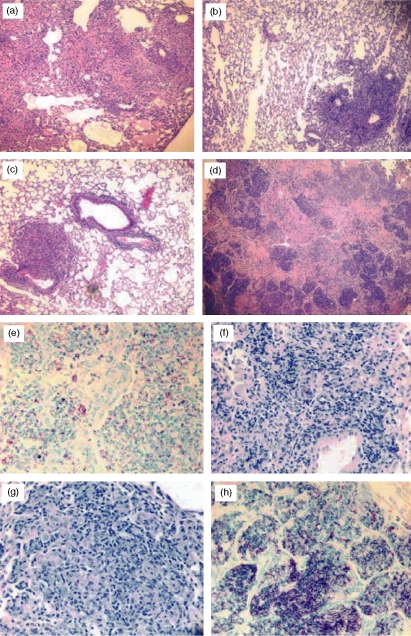
Figure 5.
Lung histopathology from the naive, vaccinated, and antibody-treated CD4–/– mice showed substantially worsening pathology only in vaccinated mice that were depleted of Thy-1.2+ cells and not in immunized CD4–/– mice that had been depleted of CD8+, TCR-γδ+ and NK1.1+ cells. Lung tissue was obtained from each group at the time of death, fixed in formalin, and embedded in paraffin. The lung sections were stained with haematoxylin & eosin (a–d) or with Ziehl–Neelsen acid-fast stain (e–h) and evaluated by light microscopy. Stained lung sections from naive (a, e), mc26030-vaccinated (b, f), mc26030-vaccinated and anti-CD8, NK1.1, and TCR-γδ mAb-treated (c, g), and mc26030-vaccinated and anti-Thy-1.2 mAb-treated (d, h) mice are shown above.

