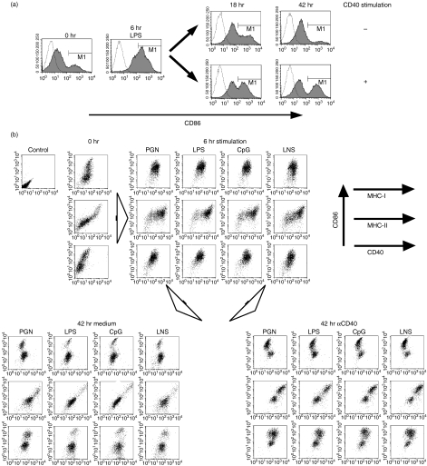
Figure 1.
Lipopolysaccharide (LPS)-induced transient activation and successive inactivation.(a) Immature dendritic cells (DCs) derived from B6 bone marrow (BM) cells were stimulated by LPS for 6 hr. The cells were then cultured with or without αCD40 for an additional 18 hr or 42 hr. Activation of DCs was analysed by fluorescence-activated cell sorter staining of anti-CD11c-FITC and anti-CD86-PE with propidium iodide live gating. Open lines indicate isotype control. (b) Immature BM-DCs of B6 mice were stimulated for 6 hr by 1 µg/ml peptidoglycan (PGN) as a toll-like receptor (TLR) 2 agonist, 1 µg/ml LPS as a TLR4 agonist, 0·1 µm cytosine-phosphorothioate-guanine (CpG) 5002 as a TLR9 agonist and lysate of necrotic B6 splenocytes (LNS), respectively. The cells were cultured for a further 42 hr with or without 5 µg/ml αCD40. The expression of Kb (MHC-I), Ab (MHC-II) and CD40 together with CD86 on BM-DCs was analysed by flow cytometry at the time-points indicated.

