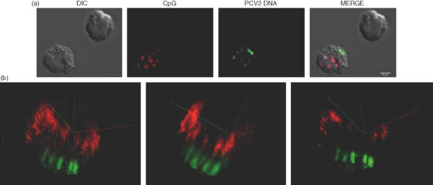
Figure 5.
Localization of cytosine-phosphorothioate-guanine oligodeoxynucleotide (CpG-ODN) and purified porcine circovirus type 2 (PCV2) DNA in natural interferon producing cells (NIPCs). Enriched NIPCs (Percoll/CD4 protocol) were incubated with 5 µg CpG-ODN CX-rhodamine-labelled (red) and 5 µg PCV2 DNA Cy5-labelled (green) for 3 hr.(a)Confocal and differential interference contrast (DIC) images of representative cells.(b)Three-dimensional views created using the colocalization software imaris.

