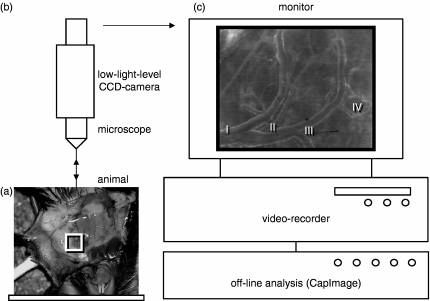
Figure 1.
Schematic illustration of the intravital microscope set-up. After preparation and eversion of the cutaneous flap at the ventral aspect of the neck, the experimental animals were placed in a supine position onto the microscope stage (a). A modified Zeiss Axiotech microscope, fixed vertically above the stage platform, was used for epi-illumination microscopy of the lymph node preparation (b). Microscopic images were televised and recorded on videotape (c) for off-line analysis of haemodynamics and leucocyte–endothelial cell adhesive interactions in individual cervical lymph node HEVs. The depicted microscopic image of the cervical lymph node microcirculation (c) shows the typical arrangement of four generations of HEVs.

