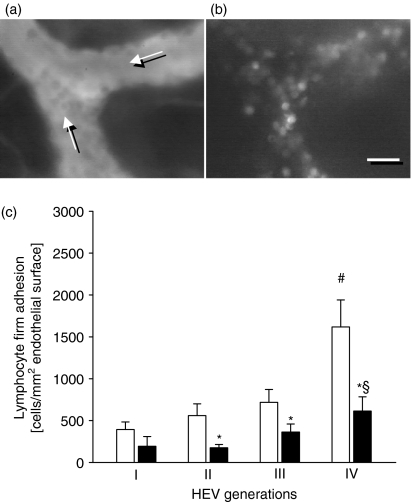
Figure 2.
Firm adhesion by lymphocytes in HEVs of the cervical lymph node is LFA-1-dependent. Intravital fluorescence microscopy of the cervical lymph node allows for visualization of individual HEVs and intravascularly adherent leucocytes, using blue-light epi-illumination and contrast enhancement of the intravascular plasma by intravenously applied fluorescein isothiocyanate-dextran (a, direction of blood flow is as indicated), as well as green-light epi-illumination and contrast enhancement by Rhodamine 6G (b), respectively. Notably, the majority of the Rhodamine 6G-stained leucocytes appear with homogeneously stained and round-shaped nuclei, indicating their lymphocytic nature. Quantitative analysis of firm lymphocyte adhesion in four generations of cervical lymph node HEVs (c) in C57BL/6 wild-type mice (white bars) and LFA-1-deficient animals (black bars) under resting conditions. Data are mean values ± SEM, n = 6 or n = 7, *P < 0·05 versus C57BL/6 wild-type mice, #P < 0·05 versus HEV generation I, §P < 0·05 versus HEV generation II. Scale bar represents 15 μm (a,b).

