Figure 2.
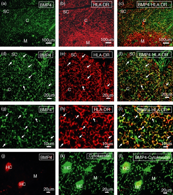
Localization of BMP4-expressing cells in the human thymus. Frozen sections of human thymus were doubled stained with anti-BMP4 (a, d, g, j) and anti-HLA-DR (b, e, h) or anti-cytokeratin (k) antibodies. (a–f) BMP4 expression (green fluorescence; a, d) is mostly restricted to the cortical and subcapsular areas, appearing as a reticular network associated with HLA-DR+ epithelial cells (red fluorescence; b, e). (g–i) BMP4 expression (green fluorescence; g) shows a punctate pattern distributed in the soma and the cellular processes of the cortical HLA-DR+ epithelial cells (red fluorescence; h). (j–l) In the thymic medulla, BMP4 expression (red fluorescence; j) is associated with cytokeratin-positive Hassall's corpuscles (HC; green fluorescence; k). Yellow fluorescence demonstrates the expression of BMP4 in subcapsular and cortical epithelial cells (c, f, i) and medullary Hassall's corpuscles (l). C, cortex; M, medulla; SC, subcapsule.

