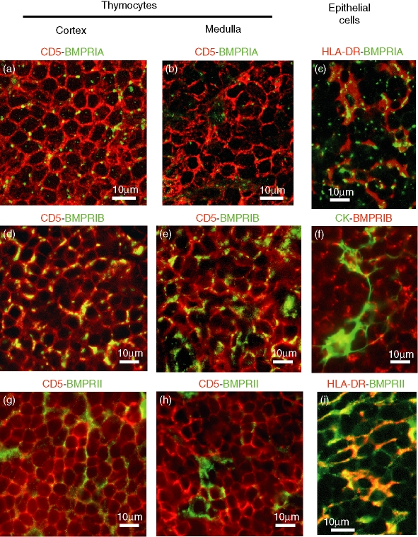
Figure 5.
Double immunofluorescence analysis of BMP2/4 receptors in human thymocytes and thymic epithelial cells. To analyse the expression of BMP receptors in thymocytes, tissue sections were double-stained with the pan-thymocyte marker CD5 (red fluorescence), and anti-BMPRIA (a, b), anti-BMPRIB (d, e) or anti-BMPRII (g, h) antibodies (all of them in green fluorescence). Yellow fluorescence demonstrates the expression of BMP receptors in thymocytes located in the thymic cortex (a, d, g) or medulla (b, e, h). Thymic sections were also double-stained with anti-HLA-DR (red fluorescence; c, i) or anti-cytokeratin (green fluorescence;f)antibodies, for the identification of thymic epithelial cells, and anti-BMPRIA (green fluorescence; c), anti-BMPRIB (red fluorescence; f) or anti-BMPRII (green fluorescence; i) antibodies. Micrographs show details of the thymic cortical area and the yellow fluorescence demonstrates BMP receptor expression in thymic epithelial cells.

