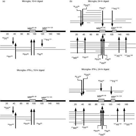
Figure 2.
Proteolytic fragments of MBP after processing with lysosomal extracts. Intact MBP was incubated with lysosomal extracts from resting microglia (microglia; top panel) or IFN-γ-stimulated microglia (microglia + IFN-γ; bottom panel) for 10 hr (a) or 24 hr (b), and the proteolytic fragments obtained were identified by mass spectrometry and displayed along with full-length MBP and the location of the two major immunogenic epitopes (MBP85–99 and MBP116–123). Arrows mark major cleavage sites and the respective flanking amino acids are indicated.

