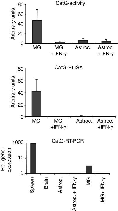
Figure 4.
Influence of IFN-γ-treatment on CatG expression and activity in microglia-derived lysosomes. Total amounts of CatG activity (top panel) and CatG protein (middle panel) were compared between equal amounts of protein from lysosomal fractions derived from resting versus IFN-γ-stimulated microglia (MG) or astrocytes (Astroc.), respectively. CatG activity was determined by turnover of the fluorogenic substrate Suc-AAPF-AMC and CatG protein by ELISA, as detailed in the materials and methods, and expressed in arbitrary units. Mean values and standard deviation of three independent experiments are presented. Bottom panel: single-cell suspensions from total spleen and total brain were compared with resting and IFN-γ-stimulated microglia and astrocytes, respectively, with regard to the expression of CatG mRNA, using quantitative PCR, as described.

