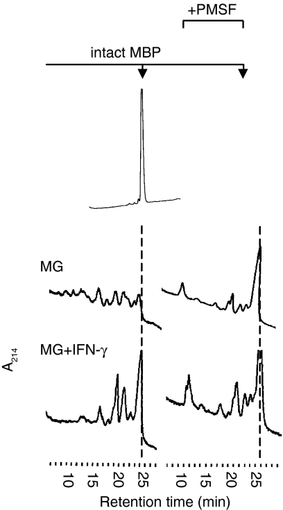
Figure 5.
PMSF treatment eliminates the effect of IFN-γ on MBP processing. Intact MBP was incubated with lysosomal extracts from resting (MG; top panels) or IFN-γ-stimulated (MG + IFN-γ; bottom panels) microglia for 24 hr, either in the absence (left panels) or presence (right panels) of the serine protease inhibitor PMSF (5 mm). Proteolytic fragments obtained are resolved by RP-HPLC and visualized by ultraviolet absorption at 214 nm (A214). The hatched line indicates the elution time of intact MBP.

