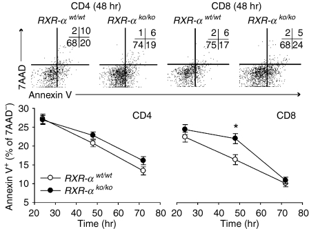
Figure 7.
Effect of Rxra disruption on CD4+ and CD8+ lymphocyte apoptosis. Data are from five wild-type (RXR-αwt/wt) and five homozygous knockout (RXR-αko/ko) mice paired by litter and gender. Cultures of splenocytes were stimulated with plate-bound anti-CD3 (10 μg/ml) and anti-CD28 (10 μg/ml). CD4+ and CD8+ lymphocytes were identified by surface staining of cells in the lymphocyte gate identified by forward-scatter and side-scatter analysis. Cells undergoing apoptosis were identified using Annexin V and dead cells were identified using 7AAD. Top panel: Annexin V and CFSE flow analysis plots of CD4+ and CD8+ cells from two representative mice 48 hr after stimulation are shown, with the number of events for each quadrant shown in the upper right-hand corner of each plot. Bottom panel: mean ± SE percentage of viable (7AAD-negative) CD4+ and CD8+ lymphocytes for all five mice of each genotype are shown at 24, 48 and 72 hr. Two-way anova was used to compare genotypes at each time-point (using duplicate measurements for each sample) while controlling for interexperiment variation. The asterisk indicates that the mean for CD8+ cells differed by genotype at 48 hr (P = 0·013).

