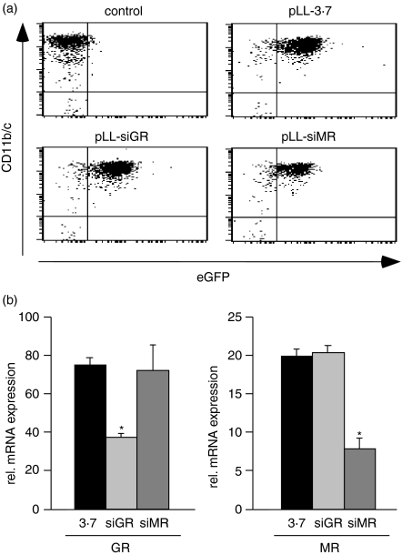
Figure 2.
(a) Flow cytometric analyses of peritoneal macrophages transduced with the lentiviruses pLL-3·7, pLL-siGR and pLL-siMR for expression of CD11b/c and eGFP. Untransduced cells are shown for comparison. (b) GR and MR mRNA expression was determined by quantitative PCR in macrophages after lentiviral gene inactivation as in (a). Statistical analysis was performed by Student's t-test. The asterisk indicates P < 0·05.

