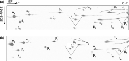
Figure 1.
Two-dimensional gels of 20S proteasomes from non-infected (a) and S. typhimurium-infected (b) B27-C1R lymphoid cells. Proteasome purifications were performed as described in the Materials and methods. About 10 μg of proteasome were analysed by two-dimensional electrophoresis, and identification of spots were performed by mass spectrometry after trypsin digestion as described in the Materials and methods.

