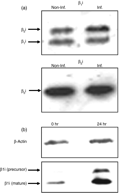
Figure 2.
Western blot analysis of proteasome subunits. (a) Western blot analysis of inducible β-subunits of the 20S proteasome from B27-C1R cells. About 2 μg purified 20S proteasomes from non-infected and infected (24 hr postinfection time) B27-C1R cells were subjected to 12% SDS-PAGE. Proteins were transferred to membranes, and analysed by Western blot. Upper panel: Western blot using the polyclonal antibody 8026.3, which recognizes the inducible subunit β1i, and cross-reacts with β5i. The intensity ratio of the β1i and the cross-reactive β5i bands between infected (Inf.) and non-infected (Non-Inf.) cells were 0·96 : 1 and 1·36 : 1, respectively. Lower panel: Western blot using the polyclonal antibody 8027.3, which recognizes the inducible subunit β5i. The intensity ratio of the bands corresponding to Inf. and Non-Inf. cells was 1·03 : 1. (b) Western blot analysis of constitutive (β1) and inducible (β1i) subunits in control and IFN-γ-induced C1R-05 cells. About 106 cells were lysed in SDS-PAGE loading buffer and subjected to 12% SDS-PAGE. Constitutive and induced subunits were detected as described in the Materials and methods. β-actin was used as a housekeeping protein. The intensity of the β1i bands, after IFN-γ stimulation was increased 2·84-fold, relative to non-stimulated cells, after normalizing to the intensity of the corresponding actin bands.

