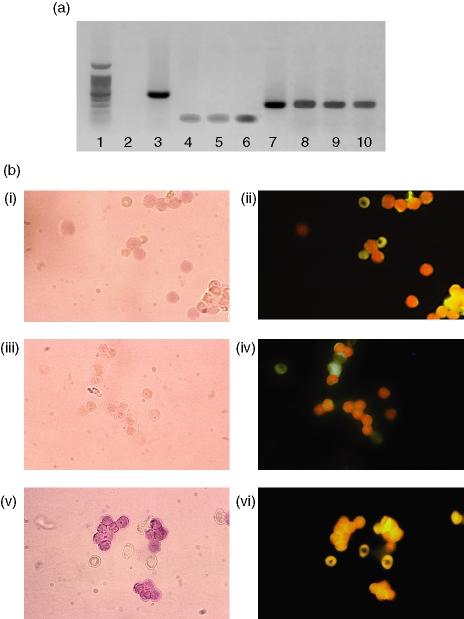
Figure 1.
Expression of 5-LOX mRNA by MOLT4 and Jurkat T cell lines as well as peripheral blood T cells. (a) Amplification of 5-LOX mRNA by RT-PCR (35 cycles). Lane 1, molecular weight ladder; lane 2, negative control – cDNA from chloramphenicol acetyl transferase mRNA with 5-LOX primers; lane 3, positive control – same cDNA with corresponding primers; lane 4, Jurkat mRNA run without reverse transcriptase; lane 5, MOLT4 mRNA run without reverse transcriptase; lane 6, T-lymphocyte mRNA run without reverse transcriptase; lane 7, 5-LOX cDNA-positive control; lane 8, Jurkat cells; lane 9, MOLT4 cells; lane 10, purified peripheral T lymphocytes. (b) Amplification of 5-LOX mRNA by RT-PCR in situ. Panel i, negative control: peripheral blood T lymphocytes submitted to RT-PCR in situ without reverse transcriptase; panel ii, same field visualized by fluorescence microscopy after incubation with anti-CD3 antibodies; panel iii, negative control: peripheral blood T lymphocytes submitted to RT-PCR in situ with primers specific for chloramphenicol acetyl transferase; panel iv, same field visualized by fluorescence microscopy after incubation with anti-CD3 antibodies, panel v, peripheral blood T lymphocytes submitted to RT-PCR in situ with specific 5-LOX primers; panel vi, same field visualized by fluorescence microscopy after incubation with anti-CD3 antibodies. Magnification 65×.

