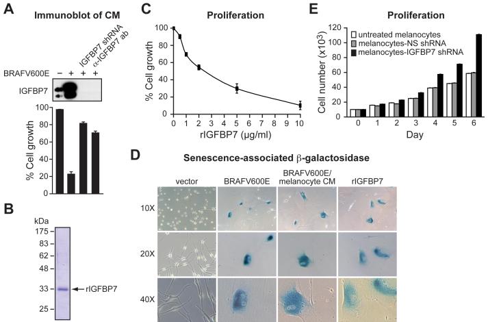
Figure 2. A Secreted Protein, IGFBP7, Induces Senescence and Apoptosis through an Autocrine/Paracrine Pathway.
(A) (Top) Immunoblot analysis of IGFBP7 levels in CM from normal melanocytes, BRAFV600E/melanocytes, BRAFV600E/melanocytes stably expressing an IGFBP7 shRNA or in BRAFV600E/melanocyte CM treated with an α-IGFBP7 antibody. (Bottom) Proliferation assays on naïve melanocytes following addition of the different CMs described above. Proliferation was measured and normalized to the growth of untreated melanocytes. Error bars represent standard error.
(B) Coomassie-stained gel of purified, recombinant IGFBP7 (rIGFBP7). Molecular weight markers are shown on the left, in kDa.
(C) Proliferation assay monitoring the effect of rIGFBP7 on the growth of melanocytes 14 days after treatment.
(D) β-galactosidase staining of melanocytes infected with a retrovirus expressing either empty vector or BRAFV600E, or melanocytes treated with CM from BRAFV600E/melanocytes or rIGFBP7. Images are shown at a magnification of 10X, 20X and 40X.
(E) Proliferation assay monitoring growth rates of untreated melanocytes, or melanocytes stably expressing an NS or IGFBP7 shRNA.

