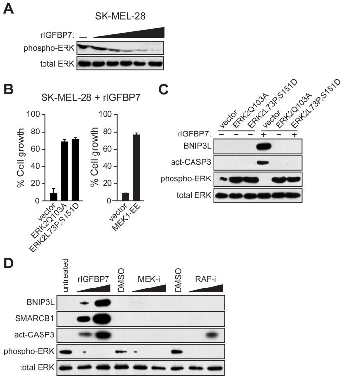
Figure 4. IGFBP7 Blocks BRAF-MEK-ERK Signaling to Activate the Apoptotic Pathway.
(A) Immunoblot analysis in SK-MEL-28 cells treated with increasing concentrations of rIGFBP7 (0.2, 1.0, 2.0, 5.0 or 10 μg/ml) for 24 hrs.
(B) Proliferation assays monitoring sensitivity of SK-MEL-28 cells to rIGFBP7. Cells were transfected with an empty expression vector or a constitutively activated ERK2 or MEK1 mutant. Cell growth was analyzed 24 hrs after treatment with rIGFBP7 and normalized to the growth of the corresponding cell line in the absence of rIGFBP7 addition. Error bars represent standard error.
(C) Immunoblot analysis in SK-MEL-28 cells stably transfected with an empty expression vector or a constitutively activated ERK2 mutant. SK-MEL-28 cells were either untreated or treated with 10 μg/ml of rIGFBP7, as indicated, for 24 hrs prior to harvesting cells.
(D) Immunoblot analysis in SK-MEL-28 cells 24 hrs following treatment with rIGFBP, a MEK inhibitor (MEK-i) or a RAF inhibitor (RAF-i).

