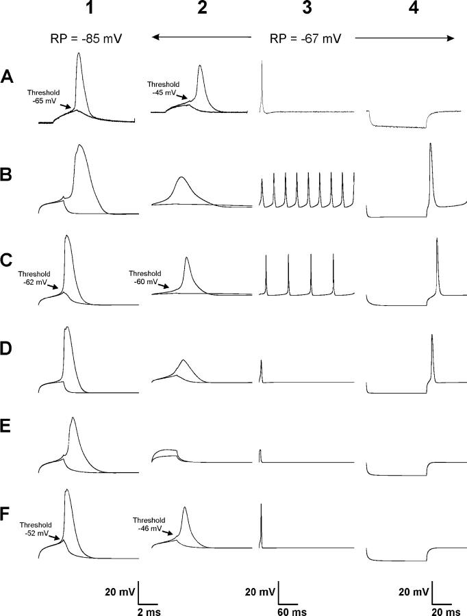
Figure 1.
Real and simulated action potentials at resting potential of −85 and −67 mV. (A) Shown are action potentials evoked at a resting potential of −85 mV (column 1, A1) and a resting potential of −67 mV in solution containing elevated extracellular K (column 2, A2). Superimposed on each action potential trace is the trace representing the largest depolarization that failed to elicit an action potential. Shown in column 3 is an action potential evoked at a resting potential of −67 mV on a long time-base (A3). In normal muscle, only a single action potential is evoked. In column 4 is a trace showing the response of the fiber to a 60-ms hyperpolarizing pulse (A4). In normal muscle, no action potential is evoked upon termination of the hyperpolarizing pulse (anode break excitation). (B) Shown are simulated action potentials that result using previously published parameters (Cannon et al., 1993). The action potentials are too wide at resting potentials of −85 and −67 mV (B1 and B2). Furthermore, a run of action potentials (myotonia) is evoked following a single depolarizing pulse at a resting potential of −67 mV (B3). Finally, there is anode break excitation following a hyperpolarizing stimulus pulse (B4). (C) After adjustment of kinetic parameters for sodium and potassium channels, simulated action potentials at a resting potential of −85 mV more closely resemble those from real fibers (C1). However, at a resting potential of −67 mV, the threshold is very close to the resting potential (C2). This results in both myotonia (C3) and anode break excitation (C4). (D) When a lower specific membrane resistance of 1,333 Ωcm2 is used, the simulated action potential at a resting potential of −85 mV looks normal (D1), but at −67 mV it is small and wide (D2). While no myotonia is present (D3), anode break excitation remains (D4). (E) When the midpoint of slow inactivation is set at −99 mV with a slope of 6 mV (Simoncini and Stuhmer, 1987; Ruff, 1996, 1999), the simulated action potential at −85 mV has a slow rise time and is somewhat wide (E1). Muscle is inexcitable at a resting potential of −67 mV (E2–4). (F) When the voltage dependence of sodium channel activation and fast inactivation are shifted by 7 mV to values of −66 and −30 mV, respectively, simulated action potentials are accurately modeled at a resting potential of −67 mV (F2). Furthermore, both myotonia and anode break excitation are prevented (F3, F4). However, at a resting potential of −85 mV, threshold for action potential initiation is too positive (F1).

