
Figure 10.
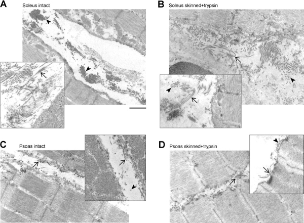
Collagen fibers in rabbit psoas and soleus. Representative electron micrographs of muscle strips before (A and C) and after (B and D) exposure to Triton X-100 (skinning) and 0.2 μg/ml trypsin (titin proteolysis), in soleus (A and B) and psoas (C and D). Note the extensive collagen depositions in intact soleus, which are barely removed during skinning. Psoas contains much less collagen than soleus. Mechanical measurements had been performed on the treated fiber bundles before preparation for EM. Arrowheads, collagen fibers in cross section; arrows, in longitudinal section. Bar, 1 μm (for all panels).