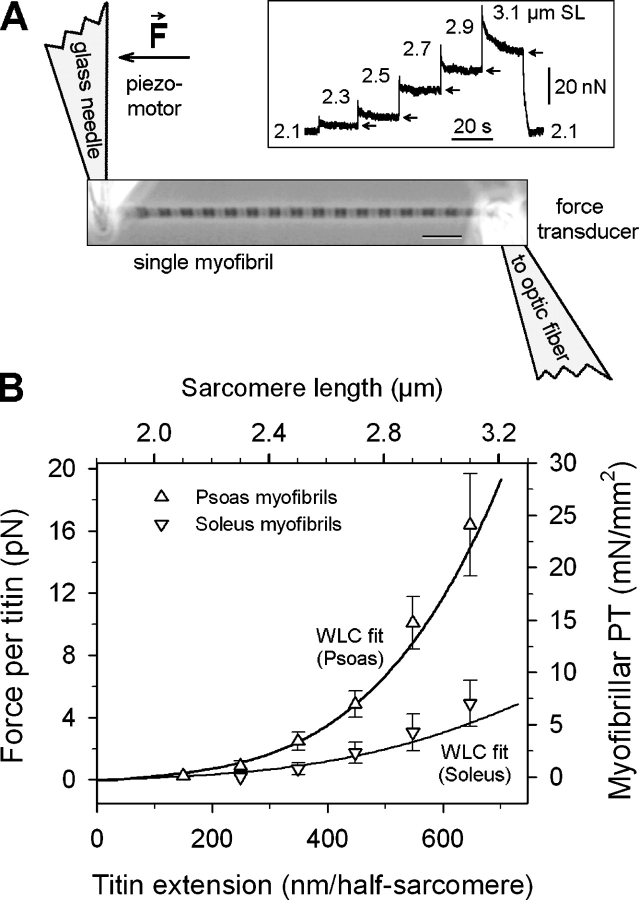
Figure 5.
Range of variability in titin-based passive force due to titin-size differences estimated by single-myofibril mechanics and wormlike chain simulations. (A) Scheme of experimental arrangement for mechanical manipulation of single myofibrils. Bar, 5 μm. Inset, typical force trace during stepwise stretch of myofibril. Arrows indicate quasi-steady-state (elastic) force. (B) Differences in titin-based force between myofibrils expressing longer titin (soleus) and those expressing short titins (psoas). Symbols (mean ± SEM) correspond to the right and top axes and indicate the elastic component of PT in single myofibrils. Lines correspond to the left and bottom axes and show wormlike chain predictions of force per titin molecule, for a muscle expressing short titins (30:70 mix of 3.4/3.3 MD isoforms, as in rabbit psoas) in comparison to a muscle expressing long titin (3.7 MD; as in human soleus or rabbit diaphragm). Titin extensions of 0–700 nm mimick the SL range 1.8–3.2 μm. The curves show the maximum and minimum forces that are to be expected from the titin size variability in rabbit muscles.

