
Figure 3.
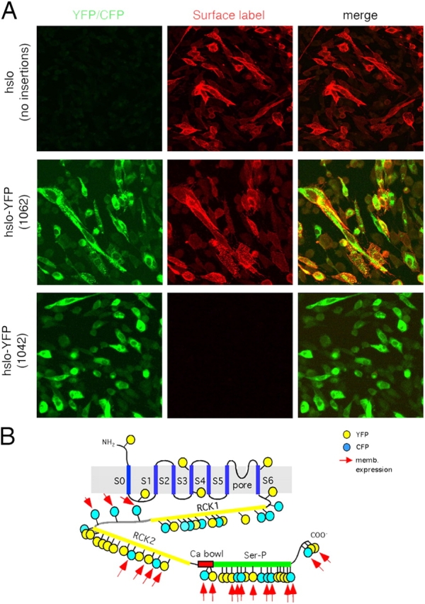
Expression of fluorescent hslo fusion proteins in the plasma membrane. (A) Confocal images of CHO cells expressing different hslo constructs. Transiently transfected hslo-YFP or CFP fusion proteins were inmunolabeled in live CHO cells before fixation. Left column, YFP fluorescence; middle column, surface immunofluorescence of hslo with Alexa 594–conjugated secondary antibody; far right, merged image. Top panels correspond to hslo channels before the transposition process. Middle and bottom panels show results obtained with two representative YFP-hslo constructs (1062-YFP in the COOH terminus and 471-YFP in the RCK1 domain). (B) 19 out of the 55 fluorescent hslo fusion proteins showed clear plasma membrane expression (red arrows). Yellow circles, YFP; blue circles, CFP. Approximate insertion sites are shown. Refer to Table I for the exact position in the hslo sequence where each insertion occurred.