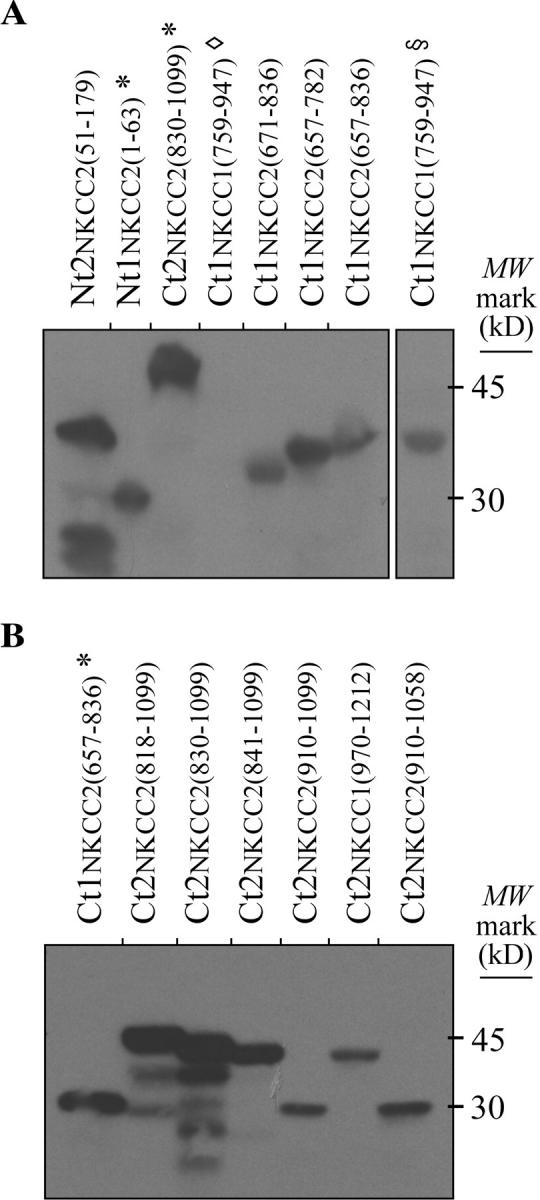
Figure 2.

Western analyses. Proteins were extracted from yeast with a lysis solution containing glass beads, 8 M urea, 40 mM Tris-HCl, 0.1 mM EDTA, 125 µM β-mercaptoethanol, 5% SDS, and protease inhibitors (final pH 6.8). In the top panel, the protein extracts were from NKCC/pGilda-transformed cells and the analyses were performed with ∼2 mg of bait proteins using an anti-LexA Ab. In the bottom panel, the protein extracts were from NKCC/pB42AD-transformed cells and the analyses were performed with ∼0.4 mg of prey proteins using an anti-HA Ab. Here, * indicates that the protein segments expressed by these transformants are currently being studied, ◊, that the protein fragment of interest had apparently degraded, and §, that the protein fragment was from another preparation of Ct1NKCC1(759–947)-transformed yeast but that it was run in a distant lane within the same gel.
