
Figure 1.
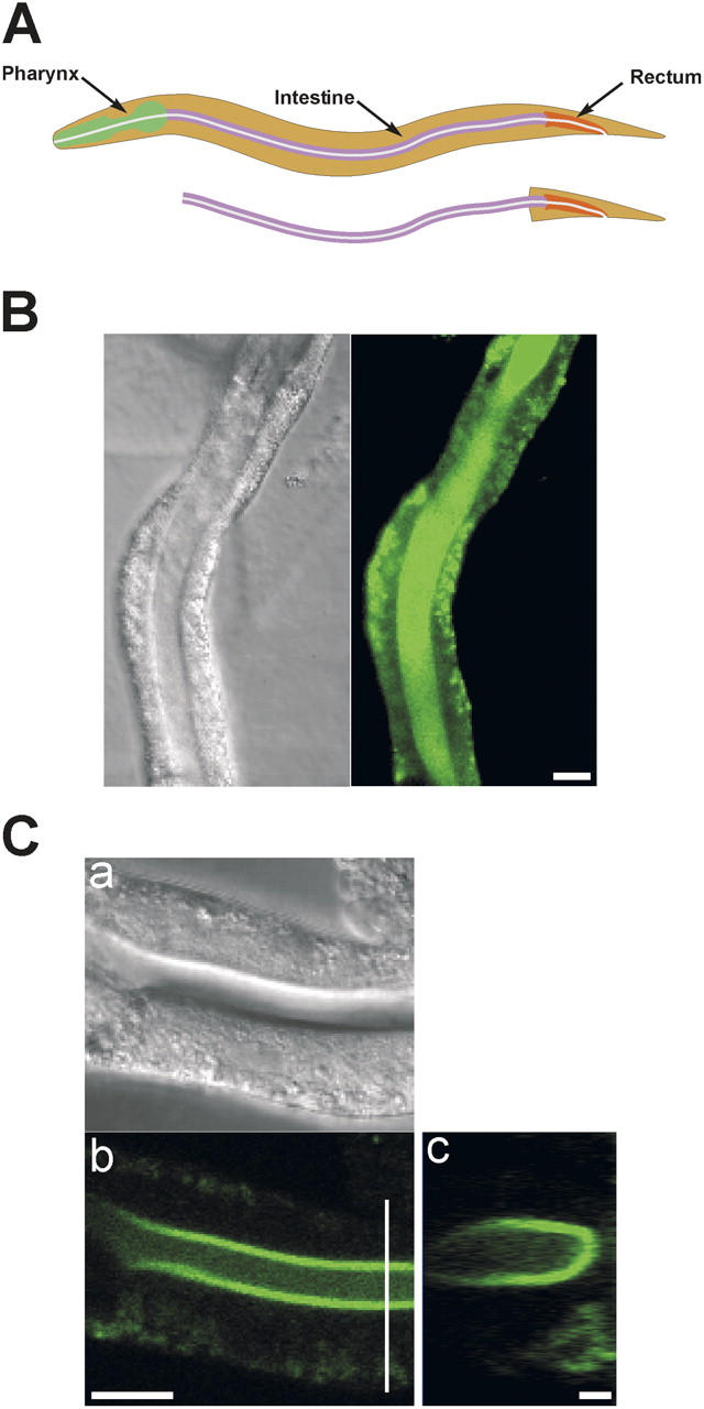
Isolated intestine preparation. (A) Schematic diagrams of worm digestive tract and isolated intestine. (B) Differential interference contrast (DIC) and fluorescence micrographs of an isolated intestine loaded with fluo-4 AM. Bar, 20 μm. (C) DIC and fluorescence confocal micrographs (panels a and b) of an isolated intestine loaded with fluo-4 AM. Panel c is a reconstruction of a series of cross sections. Location of the cross sections is shown by the white vertical line in panel b. Bars in panels b and c are 20 μm and 2 μm, respectively.