
Figure 3.
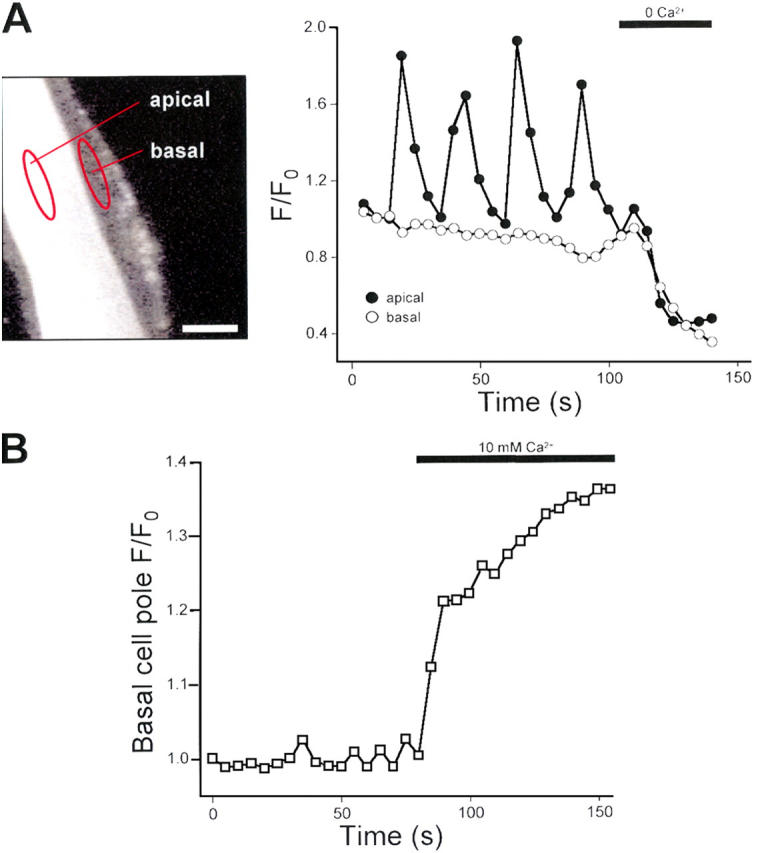
Confocal imaging of Ca2+ oscillations in isolated glo-1 intestines. (A, left) Confocal micrograph of a glo-1 intestine loaded with fluo-4 AM. Focal plane is located at the apical pole on the bottom of the intestine. Fluo-4 intensity was quantified in regions of interest outlined in red. One region is located over the apical pole of the intestine. A second region is located in the basal pole adjacent to the apical region. Bar, 10 μm. (A, right) Changes in apical and basal pole fluo-4 intensity. Calcium oscillations are detected only in the apical pole of the epithelium. Removal of extracellular Ca2+ induces similar reductions in fluo-4 intensity in both apical and basal poles. Similar results were obtained in two additional intestines. (B) Effect of elevation of bath Ca2+ on fluo-4 intensity in the basal cell pole. A region of the basal cell pole only was imaged by laser scanning. Elevation of bath Ca2+ to 10 mM induced a rapid rise in basal cell pole fluo-4 fluorescence intensity. Similar results were obtained in two additional intestines.