
Figure 9.
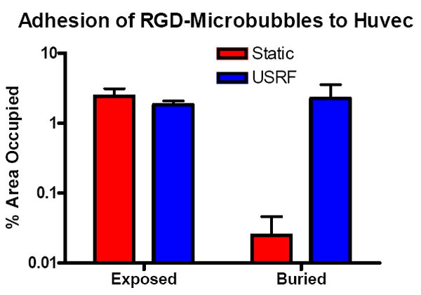
Area fractions (mean ±SD) of fluorescent microbubbles on plated Huvec cells (~50% confluence) within a circular region of interest near the center of the plate (~2-mm diameter). Shown are static adhesion results, where microbubbles were allowed to adhere owing to buoyancy force during a 5-min incubation. Also shown are USRF-assisted adhesion results, where microbubbles were pushed within the transducer focus as shown in Figure 7.