
Fig. 3.
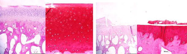
Histological sections of the impact area of OP-1 treated (left) and control sheep (right) femoral condyles 12 weeks after injury. In control sheep there is loss of most of the thickness of the cartilage due to delamination of necrotic cartilage. In OP-1-treated knees there was partial loss of the superficial zone and small fissures. H&E (×20) and safranin-O stained (×100) 5-micron-thick sections