
Fig. 1.
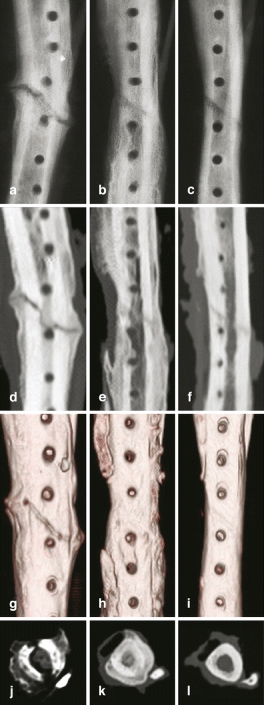
Ex vivo X-ray and computerised tomography (CT) images of canine tibia at 10 weeks post oblique osteotomy. A, B and C: X-ray images; D, E and F: 2D CT images; G, H and I: 3D CT images; J, K and L: 2D cross-sectional CT images. No rebridgement of the defect in the tibia treated with carrier alone was seen (a, d, g and j). In contrast, the tibial oblique defects treated with 1 mg (b, e, h and k) and 0.1 mg (c, f, i and l) of CP-533,536 exhibit a dose-dependent formation of new bone with full rebridgement of the defect